Ms Sharon (Ling Zhi) Heng MBBS, PhD, FRCOphth, FHEA Consultant Ophthalmic Surgeon

Rare Retina Conditions

There are numerous rare retina conditions which will need regular monitoring to optimise vision and prevent sight loss. Advanced multi modal imaging and expertise in retina conditions allows detailed monitoring and follow up of such pathologies. The following disease lists is non-exhaustive and individual diagnosis and prognosis can only be made upon consultation with Ms Heng.

Pathologic Myopia

What is pathologic Myopia or degenerative myopia

Pathologic myopia affects approximately 3% of population , in patients with high myopia, defined as refractive error of -6D or more or an axial length of 26.5mm or longer.

Pathologic myopia may result in irreversible vision loss and usually in younger or working age group.

High myopia is believed to occur due to an unusual and ongoing stretching and elongation of the eyeball, which usually begins in childhood. As a result, young individuals may find themselves in need of powerful corrective lenses early on. For those with severe myopia and significantly elongated eyes, the eyeball’s walls become excessively stretched. Consequently, all the layers that constitute the eyeball’s wall become very thin. Clinical manifestations of pathologic myopia or degenerative myopic maculopathy includes myopic choroidal neovascularization (CNVM) and CNV-related macular atrophy, diffuse chorioretinal atrophy, patchy chorioretinal atrophy, lacquer cracks or punctate inner choroidopathy (PIC)

myopic choroidal choroidal neovascularisation

Why should I monitor for pathologic Myopia

As pathologic myopia may cause vision loss, it is important to regularly monitor either yourself at home with amsler grid (looking for distortion or wavy lines) or with an ophthalmologist.

Pathologic myopia may cause the following:

  • Myopic Choroidal Neovascularization (CNV): The growth of new, weak blood vessels beneath the retina has been observed to be associated with instances of leakage or bleeding. Prompt treatment with antivegf may prevent central vision impairment.
  • Retinal detachment or tears: High myopia is well demonstrated to be a contributing factor to the development of retinal tears and detachment. Onset of floaters or flashing light will warrant urgent ophthalmic assessments.
  • Posterior staphyloma: An outward bulging of the eye wall (sclera) due to weakening and stretching. This can be monitored with imaging such as oct scans and wide field imaging.
  • Lacquer Cracks: These are fine lines or breaks in the Bruch’s membrane (an outer retina collagenous layer). These may develop into larger patches of atrophy or bleeding.
  • Glaucoma: The elevated risk of damage to the optic nerve is attributable to elevated intraocular pressure. The evaluation of glaucoma in a myopic eye may be challenging and will warrant specialised assessment due to the atypical structure of a myopic eye.
peau d’orange and angiod streaks

Angiod Streaks

Angioid streaks are bilateral, narrow, irregular lines located deep to the retina. These lines are configured in a radiating fashion, emanating from the optic disc and thought to be breaks in Bruch’s membrane ( a collagenous outer retina layer) which is thought to be weakened due to various systemic pathology.

There are several known associated systemic diseases that are associated with angioid streaks. The most prevalent of these diseases is pseudoxanthoma elasticum. Other diseases include Ehler-Danlos syndrome, Paget’s disease of bone, Sickle cell disease and other hemoglobinopathies, and sometimes, no cause can be found (idiopathic).

The diagnosis of angioid streaks is typically made on the basis of clinical observation, and optical imaging including color fundus photography or oct scans. The majority of patients are asymptomatic; however, secondary complications can result in choroidal neovascularization, causing vision loss.

Haemoglobinopathies are genetic disorders due to either abnormal haemoglobin, such as in sickle cell disease, or insufficient production of haemoglobin chains, as in thalassaemia. The most serious vision-threatening complication of sickle cell disease is proliferative sickle cell retinopathy, and this is reportedly seen in 0.5 per cent of patients with HbSS disease (the severe variant of sickle cell disease) and about 2.5 per cent of patients with HbSC disease (a less severe variant).

Hemoglobinopathy Retinopathy

Color fundus photograph showing periphery new vessels

Sickle cell retinopathy

Sickle cell retinopathy can manifest in a variety of forms, affecting the retina in different ways. sickle cell disease can cause reduced blood supply to the retina and the ischaemic retina produces an increase in a protein called vascular endothelial growth factor (VEGF) which then stimulates the growth of new blood vessels in the outer edges of the retina (peripheral retina). The newly formed blood vessels are characterised by weakness and leakage, which can result in further damage to the retina and vision.The form of retinopathy most likely to affect vision is known as proliferative sickle cell retinopathy (PSR).

The formation of new blood vessels is characterised by the formation of fan-shaped networks along the surface of the retina and the posterior of the vitreous gel within the eye.

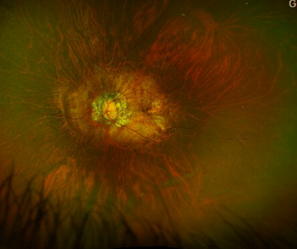
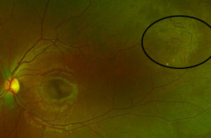
The development of these networks can vary, some may self resolve, or may cause bleeding.

In approximately 40% of cases, the new blood vessels regress, and the sea-fan resolves on its own. In other cases, the sea-fans do not exhibit significant growth and do not bleed.

Proliferative sickle cell retinopathy is more common in individuals with HbSC type of sickle disease than HbSS. Majority have be asymptomatic in early stages, but some may unfortunately have temporary or more rarely, irreversible sight loss.

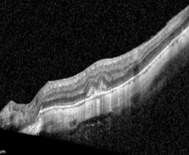
Sight loss from proliferative sickle cell retinopathy happens from vitreous haemorrhage which is a bleed from new bleed vessel into the vitreous gel or retina detachment where the retina seperates due to new blood vessel pulling on the retina (traction).

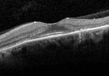
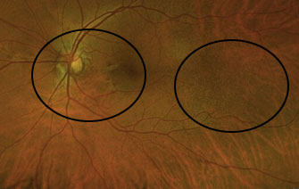
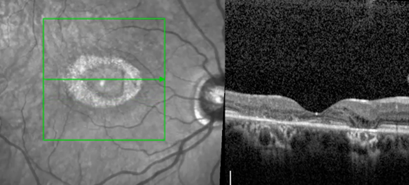
Other effects of sickle cell retinopathy include sickle cell maculopathy- where blood supply is lost in the macular or center of the retina causing demise of some of the photoreceptor cells and vision loss, retina artery occlusion where there is occlusion of the arteries delivering blood and oxygen to the retina.

Some systemic treatment or drugs for various diseases, including hydroxychloroquine for autoimmune or rheumatoid diseases or tamoxifen for breast cancer may have rare inadvertent effects on the retina. Regular monitoring is important for early diagnosis to prevent sight loss.

Drug Induced Toxic Retinopathy

Hydroxychloroquine retinotoxicity

Breast Cancer Treatment

Tamoxifen Toxicity

Tamoxifen retinopathy is a rare, dose-dependent side effect of the breast cancer drug tamoxifen. It occurs in up to 12% of patients, often with long-term use or at doses greater than 20 mg daily.

Patients may experience blurred vision and decreased visual acuity as symptoms. occasionally, there may be no symptoms at all.

Diagnosis is through ophthalmic examination and findings of characteristic white/yellow refractile deposits in the macula. OCT will show subtle cysts or hyper-reflective deposits.

Monitoring is important as the condition is irreversible and any damage is likely permanent. Management is usually to stop tamoxifen or consider alternative treatment along with patient’s oncologist, with the aim to prevent worsening of visual impairment.

Frequently Asked Questions

What is hydroxychloroquine?

Hydroxychloroquine is a major drug used for treatment and prevention of malaria

It is also used in several autoimmune diseases: rheumatoid arthritis, lupus, and porphyria cutanea tarda, Srojen syndrome. A database study published by King’s college hospital rheumatology department with 3000 patients estimates that more than 35% of their patients are on this treatment.

Recent epidemiological data indicate that the prevalence of toxicity amongst long-term ( 5 years) hydroxychloroquine users is approximately 7.5%. This risk increases to 20- 50% after 20 years, dependent on individual risk factors. In a separate US study, the prevalence of hydroxychloroquine retinopathy was found to be 0.48% in patients taking a dose of greater than 6.5mg/kg/day.

The royal college of ophthalmologists’ guidelines recommended only monitoring after 5 years of treatment, given that only 4% of patients who started hydroxychloroquine continue on it for beyond 5 years.

Factors associated with higher risks for toxicity including the following:

a. Patients on chloroquine or high dose hydroxychloroquine (>5mg/kg/day).
b. Patients with renal disease and impaired renal function (estimated glomerular filtration rate of less than 60ml/min/1.73m2)
c. Patients on Tamoxifen treatment

Previously, retinopathy was detected after patients develop symptoms by which there would have been considerable irreversible retinal pigment epithelium damage. Clinically, this has been described as a typical parafoveal pattern of RPE loss or a Bull’s eye maculopathy. This is usually associated with paracentral visual field loss and typically affects both eyes. Further, a korean study have found pericentral damage in patients of Asian ethnicity. Studies have shown continuation of worsening of visual function in patients with RPE damage for approximately 3 years even after cessation of the drug.

The advent of a plethora of imaging modality such as spectral domain OCT, widefield color fundus photography and fundus autofluorescence now allows pre-symptomatic detection of RPE damage and modifying or cessation of treatment. Early diagnosis will hence limit the progression of debilitating visual field loss. The main aims of conducting any monitoring program is not to prevent hydroxychloroquine retinopathy, but to detect the earliest definitive signs of pre-symptomatic toxicity. This will help facilitating the informed decision making process between patients and prescribing physicians on available treatment options as well as the risks and benefits. Indirectly, this will decrease the proportion of avoidable visual damage in this subgroup of patients.

OCT imaging of thining of the parafoveal region- bull’s eye maculopathy

Chloroquine is associated with a higher prevalence of toxic retinopathy when compared to hydroxychloroquine; and is used rarely in clinical practice. Guidance on safe dosing suggests a dose of less than 2.3 milligrams per kilogram per day for chloroquine. Because of the higher risk of retina toxicity, patients should be screened annually.